The green synthesis of silver nanoparticles have attracted many researchers due to their wide range of applications. The objective of this study is to synthesize silver nanoparticles using water hyacinth extract for the detection of metal ions in aquatic solutions. In the present study, the silver nanoparticles synthesis employing the leaf extract of water hyacinth as the capping and reducing agent has been reported. The particles showed absorption maxima at 406 nm establishing the formation of silver nanoparticles. The particles were characterized by FTIR, XRD, SEM-EDX, TEM and Zeta Potential. The polyphenols present in the leaf extract are accountable for reducing and the capping activity which was revealed in the FTIR spectra. XRD revealed the crystalline nature of the nanoparticles. The morphology, size and shape of the silver nanoparticles were investigated with the help of electron microscopy techniques. The silver nanoparticles are observed to be spherically shaped with an average diameter of 10.78 ± 4.61 nm. EDX spectra established the presence of elemental silver in the nanoparticles. A zeta potential of -31.7 mV was recorded indicating that the silver nanoparticles are stable. These biosynthesized silver nanoparticles were employed to detect metal ions in aqueous solutions and two metal ions (Hg2+ and Fe3+) at 1000 micro molar concentration were detected successfully. Thus, the results of the study indicate that the silver nanoparticles synthesized from water hyacinth leaf extract have potential application in the detection of metal ions.
| Published in | American Journal of Nano Research and Applications (Volume 13, Issue 1) |
| DOI | 10.11648/j.nano.20251301.12 |
| Page(s) | 16-27 |
| Creative Commons |
This is an Open Access article, distributed under the terms of the Creative Commons Attribution 4.0 International License (http://creativecommons.org/licenses/by/4.0/), which permits unrestricted use, distribution and reproduction in any medium or format, provided the original work is properly cited. |
| Copyright |
Copyright © The Author(s), 2025. Published by Science Publishing Group |
Silver Nanoparticles, Water Hyacinth, Green Synthesis, Metal Detection, Heavy Metals
Plant part used | Plant | Heavy metals detected | References |
|---|---|---|---|
Fruits | Water apple (Syzygium aqueum) | Hg2+ | [63] |
Watermelon (Citrullus lanatus) | Hg2+, Cu2+ | [73] | |
Kokum fruit | Hg2+ | [74] | |
Carica papaya | Hg2+, Fe3+ | [75] | |
Flower | Acacia nilotica | Hg2+ | [62] |
Moringa oleifera | Cu4+ | [76] | |
Anchusa azurea | Hg2+ | [77] | |
Roots | Bistorta amplexicaulis | Hg2+, Pb2+ | [78] |
Panax ginseng | Hg2+ | [79] | |
Soymida febrifuga | Hg2+ | [80] | |
Peel | Allium cepa L | Hg2+ | [81] |
Sapota (Manilkara zapota L.) | Hg2+, Co2+ | [82] | |
Leaves | Cordia myxa | Hg2+, Fe3+ | [51] |
Trigonella foenum-graecum L. | Hg2+, Fe3+ | [65] | |
Sonchus arvensis L | Hg2+, Fe3+ | [66] | |
Dahlia pinnata | Hg2+ | [70] | |
Artemisia vulgaris | Hg2+ | [83] | |
Yemeni Mistletoe (Phragmanthera austroarabica) | Cr (VI) | [84] | |
Acacia chundra | Hg2+ | [85] | |
Acalypha hispida | Mn2+ | [86] | |
Lantana camara | Hg2+, Cu2+, Pb2+, Mn2+ | [87] |
FTIR | Fourier Transform InfraRed Spectroscopy |
XRD | X-ray Diffraction |
SEM-EDX | Scanning Electron Microscopy - Energy Dispersive X-Ray Spectroscopy |
TEM | Transmission Electron Microscopy |
Ag-NPs | Silver Nanoparticles |
UV | Ultraviolet |
ICPMS | Inductively Coupled Plasma Mass Spectrometry |
ICP-OES | Inductively Coupled Plasma Optical Emission Spectroscopy |
AFS | Atomic Fluorescence Spectroscopy |
HPLC | High Performance Liquid Chromatography |
FAAS | Flame Atomic Absorption Spectroscopy |
| [1] | Narayan, N., Meiyazhagan, A., Vajtai, R. Metal nanoparticles as green catalysts. Materials. 2019, 12(21), p. 3602. |
| [2] | Palomo, J. M. Filice, M. Biosynthesis of metal nanoparticles: novel efficient heterogeneous nanocatalysts. Nanomaterials. 2016, 6(5), p. 84. |
| [3] | Hu, H., Xin, J. H., Hu, H., Wang, X., Miao, D., Liu, Y. Synthesis and stabilization of metal nanocatalysts for reduction reactions–a review. Journal of materials chemistry A. 2015, 3(21), pp. 11157-11182. |
| [4] | Wang, Y., Yan, B., Chen, L. SERS tags: novel optical nanoprobes for bioanalysis. Chemical reviews. 2013, 113(3), pp. 1391-1428. |
| [5] | Luo, C., Wang, Y., Li, X., Jiang, X., Gao, P., Sun, K., Zhou, J., Zhang, Z., Jiang, Q. An optical sensor with polyaniline-gold hybrid nanostructures for monitoring pH in saliva. Nanomaterials. 2017, 7(3), p. 67. |
| [6] | Giraldo, J. P., Wu, H., Newkirk, G. M., Kruss, S. Nanobiotechnology approaches for engineering smart plant sensors. Nature nanotechnology. 2019, 14(6), pp. 541-553. |
| [7] | Burratti, L., Bolli, E., Casalboni, M., De Matteis, F., Mochi, F., Francini, R., Casciardi, S., Prosposito, P. Synthesis of fluorescent ag nanoclusters for sensing and imaging applications. In Materials Science Forum. 2018, Vol. 941, pp. 2243-2248. Trans Tech Publications Ltd. |
| [8] | Saravanan, S., Kato, R., Balamurugan, M., Kaushik, S., Soga, T. Efficiency improvement in dye sensitized solar cells by the plasmonic effect of green synthesized silver nanoparticles. Journal of Science: Advanced Materials and Devices. 2017, 2(4), pp. 418-424. |
| [9] | Venditti, I. Morphologies and functionalities of polymeric nanocarriers as chemical tools for drug delivery: A review. Journal of King Saud University-Science. 2019, 31(3), pp. 398-411. |
| [10] | Fratoddi, I., Venditti, I., Battocchio, C., Carlini, L., Amatori, S., Porchia, M., Tisato, F., Bondino, F., Magnano, E., Pellei, M., Santini, C. Highly hydrophilic gold nanoparticles as carrier for anticancer copper (I) complexes: Loading and release studies for biomedical applications. Nanomaterials. 2019, 9(5), p. 772. |
| [11] | Wei, L., Lu, J., Xu, H., Patel, A., Chen, Z. S., Chen, G. Silver nanoparticles: synthesis, properties, and therapeutic applications. Drug discovery today. 2015, 20(5), pp. 595-601. |
| [12] | Barkalina, N., Charalambous, C., Jones, C., Coward, K. Nanotechnology in reproductive medicine: emerging applications of nanomaterials. Nanomedicine: Nanotechnology, Biology and Medicine. 2014, 10(5), pp. e921-e938. |
| [13] | Singh, T., Singh, A., Wang, W., Yadav, D., Kumar, A., Singh, P. K. Biosynthesized nanoparticles and its implications in agriculture. In Biological Synthesis of Nanoparticles and Their Applications. 2019, pp. 257-274. CRC Press. ISBN-13: 978-0-367-21069-4 |
| [14] | Ashrafi, G., Nasrollahzadeh, M., Jaleh, B., Sajjadi, M., Ghafuri, H. Biowaste-and nature-derived (nano) materials: Biosynthesis, stability and environmental applications. Advances in Colloid and Interface Science. 2022, p. 102599. |
| [15] | Ahmed, S. F., Mofijur, M., Rafa, N., Chowdhury, A. T., Chowdhury, S., Nahrin, M., Islam, A. S., Ong, H. C. Green approaches in synthesising nanomaterials for environmental nanobioremediation: Technological advancements, applications, benefits and challenges. Environmental Research. 2022, 204, p. 111967. |
| [16] | Corsi, I., Winther-Nielsen, M., Sethi, R., Punta, C., Della Torre, C., Libralato, G., Lofrano, G., Sabatini, L., Aiello, M., Fiordi, L., Cinuzzi, F. Ecofriendly nanotechnologies and nanomaterials for environmental applications: Key issue and consensus recommendations for sustainable and ecosafe nanoremediation. Ecotoxicology and Environmental Safety. 2018, 154, pp. 237-244. |
| [17] | Mehta, M., Sharma, M., Pathania, K., Jena, P. K., Bhushan, I. Degradation of synthetic dyes using nanoparticles: a mini-review. Environmental Science and Pollution Research. 2021, 28(36), pp. 49434-49446. |
| [18] | Pasha, A., Kumbhakar, D. V., Sana, S. S., Ravinder, D., Lakshmi, B. V., Kalangi, S. K., Pawar, S. C. Role of Biosynthesized Ag-NPs Using Aspergillus niger (MK503444. 1) in Antimicrobial, Anti-Cancer and Anti-Angiogenic Activities. Frontiers in Pharmacology. 2021, 12. |
| [19] | Zhang, X. F., Liu, Z. G., Shen, W., Gurunathan, S. Silver nanoparticles: synthesis, characterization, properties, applications, and therapeutic approaches. International journal of molecular sciences. 2016, 17(9), p. 1534. |
| [20] | Nile, S. H., Baskar, V., Selvaraj, D., Nile, A., Xiao, J., Kai, G. Nanotechnologies in food science: applications, recent trends, and future perspectives. Nano-micro letters. 2020, 12(1), pp. 1-34. |
| [21] | Francis, S., Joseph, S., Koshy, E. P., Mathew, B. Microwave assisted green synthesis of silver nanoparticles using leaf extract of elephantopus scaber and its environmental and biological applications. Artificial cells, nanomedicine, and biotechnology. 2018, 46(4), pp. 795-804. |
| [22] | Tamilselvan, S., Soniya, R. M., Vasantharaja, R., Kannan, M., Supriya, S., Batvari, B. P. D., Ramesh, T., Govindaraju, K. Silver nanoparticles based spectroscopic sensing of eight metal ions in aqueous solutions. Environmental Research. 2022, p. 113585. |
| [23] | Devi, H. S., Boda, M. A., Shah, M. A., Parveen, S., Wani, A. H. Green synthesis of iron oxide nanoparticles using Platanus orientalis leaf extract for antifungal activity. Green Processing and Synthesis. 2019, 8(1), pp. 38-45. |
| [24] | Alsammarraie, F. K., Wang, W., Zhou, P., Mustapha, A., Lin, M. Green synthesis of silver nanoparticles using turmeric extracts and investigation of their antibacterial activities. Colloids and Surfaces B: Biointerfaces. 2018, 171, pp. 398-405. |
| [25] | Kataria, N., Garg, V. K. Green synthesis of Fe3O4 nanoparticles loaded sawdust carbon for cadmium (II) removal from water: regeneration and mechanism. Chemosphere. 2018, 208, pp. 818-828. |
| [26] | Nasrollahzadeh, M., Sajadi, S. M. Pd nanoparticles synthesized in situ with the use of Euphorbia granulate leaf extract: Catalytic properties of the resulting particles. Journal of colloid and interface science. 2016, 462, pp. 243-251. |
| [27] | Huq, M. A. Green synthesis of silver nanoparticles using Pseudoduganella eburnea MAHUQ-39 and their antimicrobial mechanisms investigation against drug resistant human pathogens. International journal of molecular sciences. 2020, 21(4), p. 1510. |
| [28] | Tyagi, S., Tyagi, P. K., Gola, D., Chauhan, N., Bharti, R. K. Extracellular synthesis of silver nanoparticles using entomopathogenic fungus: characterization and antibacterial potential. SN Applied Sciences. 2019, 1(12), pp. 1-9. |
| [29] | Lomelí-Rosales, D. A., Zamudio-Ojeda, A., Reyes-Maldonado, O. K., López-Reyes, M. E., Basulto-Padilla, G. C., Lopez-Naranjo, E. J., Zuñiga-Mayo, V. M., Velázquez-Juárez, G. Green Synthesis of Gold and Silver Nanoparticles Using Leaf Extract of Capsicum chinense Plant. Molecules. 2022, 27(5), p. 1692. |
| [30] | Bharadwaj, K. K., Rabha, B., Pati, S., Choudhury, B. K., Sarkar, T., Gogoi, S. K., Kakati, N., Baishya, D., Kari, Z. A., Edinur, H. A. Green synthesis of silver nanoparticles using Diospyros malabarica fruit extract and assessments of their antimicrobial, anticancer and catalytic reduction of 4-nitrophenol (4-NP). Nanomaterials. 2021, 11(8), p. 1999. |
| [31] | Baran, A., Keskin, C., Baran, M. F., Huseynova, I., Khalilov, R., Eftekhari, A., Irtegun-Kandemir, S., Kavak, D. E. Ecofriendly synthesis of silver nanoparticles using ananas comosus fruit peels: anticancer and antimicrobial activities. Bioinorganic Chemistry and Applications. 2021. |
| [32] | Elangovan, M., Ramachandran, D., Rajesh, K. Green Synthesis of Silver Nanoparticles Using Flower Extract of Hemigraphis colorata as Reducing Agent and its Biological Activity. Lett. Appl. NanoBioScience. 2021, 10, pp. 2646-2654. |
| [33] | Nawabjohn, M. S., Sivaprakasam, P., Anandasadagopan, S. K., Begum, A. A., Pandurangan, A. K. Green synthesis and characterisation of silver nanoparticles using Cassia tora seed extract and investigation of antibacterial potential. Applied Biochemistry and Biotechnology. 2021, pp. 1-15. |
| [34] | Pungle, R., Nile, S. H., Makwana, N., Singh, R., Singh, R. P., Kharat, A. S. Green synthesis of silver nanoparticles using the Tridax Procumbens plant extract and screening of its antimicrobial and anticancer activities. Oxidative Medicine and Cellular Longevity. 2022, (1), p. 9671594. |
| [35] | Rafique, M., Sadaf, I., Rafique, M. S., Tahir, M. B. A review on green synthesis of silver nanoparticles and their applications. Artificial cells, nanomedicine, and biotechnology. 2017, 45(7), pp. 1272-1291. |
| [36] | Ahmad, S., Munir, S., Zeb, N., Ullah, A., Khan, B., Ali, J., Bilal, M., Omer, M., Alamzeb, M., Salman, S. M., Ali, S. Green nanotechnology: A review on green synthesis of silver nanoparticles—An ecofriendly approach. International journal of nanomedicine. 2019, 14, p. 5087. |
| [37] | Vanlalveni, C., Lallianrawna, S., Biswas, A., Selvaraj, M., Changmai, B., Rokhum, S. L. Green synthesis of silver nanoparticles using plant extracts and their antimicrobial activities: A review of recent literature. RSC advances. 2021, 11(5), pp. 2804-2837. |
| [38] | Zaynab, M., Al-Yahyai, R., Ameen, A., Sharif, Y., Ali, L., Fatima, M., Khan, K. A., Li, S. Health and environmental effects of heavy metals. Journal of King Saud University-Science. 2022, 34(1), p. 101653. |
| [39] |
Jaiswal, A., Verma, A., Jaiswal, P. Detrimental effects of heavy metals in soil, plants, and aquatic ecosystems and in humans. Journal of Environmental Pathology, Toxicology and Oncology. 2018, 37(3).
https://doi.org/10.1615/JEnvironPatholToxicolOncol.2018025348 |
| [40] | Prosposito, P., Burratti, L., Venditti, I. Silver nanoparticles as colorimetric sensors for water pollutants. Chemosensors. 2020, 8(2), p. 26. |
| [41] | Jarujamrus, P., Amatatongchai, M., Thima, A., Khongrangdee, T., Mongkontong, C. Selective colorimetric sensors based on the monitoring of an unmodified silver nanoparticles (AgNPs) reduction for a simple and rapid determination of mercury. Spectrochimica Acta Part A: Molecular and Biomolecular Spectroscopy. 2015, 142, pp. 86-93. |
| [42] | Sabela, M., Balme, S., Bechelany, M., Janot, J. M., Bisetty, K. A review of gold and silver nanoparticle‐based colorimetric sensing assays. Advanced Engineering Materials. 2017, 19(12), p. 1700270. |
| [43] | Alberti, G., Zanoni, C., Magnaghi, L. R., Biesuz, R. Gold and silver nanoparticle-based colorimetric sensors: new trends and applications. Chemosensors. 2021, 9(11), p. 305. |
| [44] | Malik, A. Environmental challenge vis a vis opportunity: the case of water hyacinth. Environment international. 2007, 33(1), pp. 122-138. |
| [45] | Hublikar, L. V., Ganachari, S. V., Raghavendra, N., Patil, V. B., Banapurmath, N. R. Green synthesis silver nanoparticles via Eichhornia Crassipes leaves extract and their applications. Current Research in Green and Sustainable Chemistry. 2021, 4, p. 100212. |
| [46] | Nandiyanto, A. B. D., Ragadhita, R., Hofifah, S. N., Al Husaeni, D. F., Al Husaeni, D. N., Fiandini, M., Luckiardi, S., Soegoto, E. S., Darmawan, A. and Aziz, M. Progress in the utilization of water hyacinth as effective biomass material. Environment, Development and Sustainability. 2024, 26(10), pp. 24521-24568. |
| [47] | Oluwafemi, O. S., Anyik, J. L. and Zikalala, N. E. Biosynthesis of silver nanoparticles from water hyacinth plant leaves extract for colourimetric sensing of heavy metals. Nano-Structures & Nano-Objects. 2019, 20, p. 100387. |
| [48] | Chutrakulwong, F., Thamaphat, K. and Intarasawang, M. Investigating UV-Irradiation Parameters in the Green Synthesis of Silver Nanoparticles from Water Hyacinth Leaf Extract: Optimization for Future Sensor Applications. Nanomaterials. 2024, 14(12), p. 1018. |
| [49] | Martínez-Espinosa, J. C., Ramírez-Morales, M. A., Carrera-Cerritos, R. Silver Nanoparticles Synthesized Using Eichhornia crassipes Extract from Yuriria Lagoon, and the Perspective for Application as Antimicrobial Agent. Crystals. 2022, 12(6), p. 814. |
| [50] | Sastry, M., Mayya, K. S., Bandyopadhyay, K. pH Dependent changes in the optical properties of carboxylic acid derivatized silver colloidal particles. Colloids and Surfaces A: Physicochemical and Engineering Aspects. 1997, 127(1-3), pp. 221-228. |
| [51] | Azimpanah, R., Solati, Z., Hashemi, M. Green synthesis of silver nanoparticles and their applications as colorimetric probe for determination of Fe3+ and Hg2+ ions. IET nanobiotechnology. 2018, 12(5), pp. 673-677. |
| [52] | Patil, R. B. and Chougale, A. D. Analytical methods for the identification and characterization of silver nanoparticles: A brief review. Materials Today: Proceedings. 2021, 47, pp. 5520-5532. |
| [53] | Prakash, P., Gnanaprakasam, P., Emmanuel, R., Arokiyaraj, S., Saravanan, M. Green synthesis of silver nanoparticles from leaf extract of Mimusops elengi, Linn. for enhanced antibacterial activity against multi drug resistant clinical isolates. Colloids and Surfaces B: Biointerfaces. 2013, 108, pp. 255-259. |
| [54] | Jemal, K., Sandeep, B. V., Pola, S. Synthesis, characterization, and evaluation of the antibacterial activity of Allophylus serratus leaf and leaf derived callus extracts mediated silver nanoparticles. Journal of Nanomaterials. 2017, (1), p. 4213275. |
| [55] | Roy, K., Sarkar, C. K., Ghosh, C. K. Plant-mediated synthesis of silver nanoparticles using parsley (Petroselinum crispum) leaf extract: spectral analysis of the particles and antibacterial study. Applied Nanoscience. 2015(b), 5(8), pp. 945-951. |
| [56] | Beldjilali, M., Mekhissi, K., Khane, Y., Chaibi, W., Belarbi, L., Bousalem, S. Antibacterial and antifungal efficacy of silver nanoparticles biosynthesized using leaf extract of Thymus algeriensis. Journal of Inorganic and Organometallic Polymers and Materials. 2020, 30(6), pp. 2126-2133. |
| [57] | Edison, T. N. J. I., Atchudan, R., Sethuraman, M. G., Lee, Y. R. Reductive-degradation of carcinogenic azo dyes using Anacardium occidentale testa derived silver nanoparticles. Journal of Photochemistry and Photobiology B: Biology. 2016, 162, pp. 604-610. |
| [58] | Dos Santos, K. C., da Silva, M. F. G., Pereira-Filho, E. R., Fernandes, J. B., Polikarpov, I., Forim, M. R. Polymeric nanoparticles loaded with the 3, 5, 3′-triiodothyroacetic acid (Triac), a thyroid hormone: factorial design, characterization, and release kinetics. Nanotechnology, science and applications. 2012, pp. 37-48. |
| [59] | Ramanathan, S., Gopinath, S. C., Anbu, P., Lakshmipriya, T., Kasim, F. H., Lee, C. G. Eco-friendly synthesis of Solanum trilobatum extract-capped silver nanoparticles is compatible with good antimicrobial activities. Journal of Molecular Structure. 2018, 1160, pp. 80-91. |
| [60] | Deng, L., Ouyang, X., Jin, J., Ma, C., Jiang, Y., Zheng, J., Li, J., Li, Y., Tan, W., Yang, R. Exploiting the higher specificity of silver amalgamation: selective detection of mercury (II) by forming Ag/Hg amalgam. Analytical chemistry. 2013, 85(18), pp. 8594-8600. |
| [61] | Awad, M. A., Hendi, A. A., Ortashi, K. M., Alzahrani, B., Soliman, D., Alanazi, A., Alenazi, W., Taha, R. M., Ramadan, R., El-Tohamy, M., AlMasoud, N. Biogenic synthesis of silver nanoparticles using Trigonella foenum-graecum seed extract: Characterization, photocatalytic and antibacterial activities. Sensors and Actuators A: Physical. 2021 323, p. 112670. |
| [62] | Kahandal, A. W., Sharma, L., Sirdeshmukh, V., Kulkarni, A., Tagad, C. K. A sensitive image-based optical detection of heavy metal ions using green synthesized silver nanoparticles. International Journal of Environmental Science and Technology. 2023, 20(8), pp. 9077-9088. |
| [63] | Firdaus, M. L., Fitriani, I., Wyantuti, S., Hartati, Y. W., Khaydarov, R., Mcalister, J. A., Obata, H., Gamo, T. Colorimetric detection of mercury (II) ion in aqueous solution using silver nanoparticles. Analytical Sciences. 2017, 33(7), pp. 831-837. |
| [64] | Zou, R., Guo, X., Yang, J., Li, D., Peng, F., Zhang, L., Wang, H. and Yu, H. Selective etching of gold nanorods by ferric chloride at room temperature. CrystEngComm. 2009, 11(12), pp. 2797-2803. |
| [65] | Moond, M., Singh, S., Sangwan, S., Devi, P., Beniwal, A., Rani, J., Kumari, A. and Rani, S. Biosynthesis of silver nanoparticles utilizing leaf extract of Trigonella foenum-graecum L. for catalytic dyes degradation and colorimetric sensing of Fe3+/Hg2+. Molecules. 2023, 28(3), p. 951. |
| [66] | Chandraker, S. K., Ghosh, M. K., Lal, M., Ghorai, T. K., Shukla, R. Colorimetric sensing of Fe 3+ and Hg 2+ and photocatalytic activity of green synthesized silver nanoparticles from the leaf extract of Sonchus arvensis L. New Journal of Chemistry. 2019, 43(46), pp. 18175-18183. |
| [67] | Dayanidhi, K., Eusuff, N. S. Distinctive detection of Fe 2+ and Fe 3+ by biosurfactant capped silver nanoparticles via naked eye colorimetric sensing. New Journal of Chemistry. 2021, 45(22), pp. 9936-9943. |
| [68] | Ankamwar, B. Non-Aggregation Based Colorimetric Detection of Cu (II) and Fe (III) Using Biosynthesized Silver Nanoparticles. Chem Sci Rev Lett. 2016, 5(19), 326-330. |
| [69] | Ebrahimi, A., Samari, F., Eftekhar, E., Yousefinejad, S. Rapid and efficient colorimetric sensing of clindamycin and Fe3+ using controllable phyto-synthesized silver/silver chloride nanoparticles by Syzygium cumini fruit extract. Journal of Analytical Science and Technology. 2022, 13(1), pp. 1-16. |
| [70] | Roy, K., Sarkar, C. K., Ghosh, C. K. Rapid colorimetric detection of Hg2+ ion by green silver nanoparticles synthesized using Dahlia pinnata leaf extract. Green Processing and Synthesis. 2015, 4(6), pp. 455-461. |
| [71] | Zou, Y., Pang, J., Zhang, F., Chai, F. Silver Nanoparticles for Colorimetric Detection and Discrimination of Mercury Ions in Lake Water. ChemistrySelect. 2021, 6(24), pp. 6077-6082. |
| [72] | Farhadi, K., Forough, M., Molaei, R., Hajizadeh, S., Rafipour, A. Highly selective Hg2+ colorimetric sensor using green synthesized and unmodified silver nanoparticles. Sensors and Actuators B: Chemical. 2012, 161(1), pp. 880-885. |
| [73] | Maiti, S., Barman, G., Konar Laha, J. Detection of heavy metals (Cu+ 2, Hg+ 2) by biosynthesized silver nanoparticles. Applied Nanoscience. 2016, 6, pp. 529-538. |
| [74] | Sangaonkar, G. M., Desai, M. P., Dongale, T. D., Pawar, K. D. Selective interaction between phytomediated anionic silver nanoparticles and mercury leading to amalgam formation enables highly sensitive, colorimetric and memristor-based detection of mercury. Scientific Reports. 2020, 10(1), p. 2037. |
| [75] | Revathi, E., Yaku, G., Unnisa, S. A., Malyala, P., Praveen, V. Microwave assisted green synthesis of silver nanoparticles from Carica papaya fruit extract: Characterization and detection of Fe3+ and Hg2+ ions. Materials Today: Proceedings. 2023, 92, pp. 490-497. |
| [76] | Bindhu, M. R., Umadevi, M., Esmail, G. A., Al-Dhabi, N. A., Arasu, M. V. Green synthesis and characterization of silver nanoparticles from Moringa oleifera flower and assessment of antimicrobial and sensing properties. Journal of Photochemistry and Photobiology B: Biology. 2020, 205, p. 111836. |
| [77] | Ödemiş, Ö., Salih Ağırtaş, M. Environmentally friendly use of silver nanoparticles prepared with Anchusa azurea flower to detect mercury (II) ions. Materials Research Innovations. 2024, 28(7), pp. 555–562. |
| [78] | Ahmed, F., Kabir, H., Xiong, H. Dual colorimetric sensor for Hg2+/Pb2+ and an efficient catalyst based on silver nanoparticles mediating by the root extract of Bistorta amplexicaulis. Frontiers in Chemistry. 2020, 8, p. 591958. |
| [79] | Tagad, C., Seo, H. H., Tongaonkar, R., Yu, Y. W., Lee, J. H., Dingre, M., Kulkarni, A., Fouad, H., Ansari, S. A., Moh, S. H. Green Synthesis of Silver Nanoparticles Using Panax Ginseng Root Extract for the Detection of Hg2+. Sensors & Materials. 2017, 29(2). |
| [80] | Sowmyya, T., Lakshmi, G. V. Soymida febrifuga aqueous root extract maneuvered silver nanoparticles as mercury nanosensor and potential microbicide. World Scientific News. 2018, 114, pp. 84-105. |
| [81] | Santhosh, A., Theertha, V., Prakash, P., Chandran, S. S. From waste to a value added product: Green synthesis of silver nanoparticles from onion peels together with its diverse applications. Materials Today: Proceedings. 2021, 46, pp. 4460-4463. |
| [82] | Beniwal, A., Singh, S., Rani, J., Moond, M., Kakkar, S., Sangwan, S., Kumari, S. Waste upcycling of Sapota peels as a green route for the synthesis of silver nanoparticles and their application as catalytic and colorimetric detection of Co2+ and Hg2+. Discover Nano. 2024, 19(1), p. 191. |
| [83] | Adhikari, A., Lamichhane, L., Adhikari, A., Gyawali, G., Acharya, D., Baral, E. R., Chhetri, K. Green synthesis of silver nanoparticles using Artemisia vulgaris extract and its application toward catalytic and metal-sensing activity. Inorganics. 2022, 10(8), p. 113. |
| [84] | Alahdal, F. A., Qashqoosh, M. T., Manea, Y. K., Salem, M. A., Khan, R. H., Naqvi, S. Ultrafast fluorescent detection of hexavalent chromium ions, catalytic efficacy and antioxidant activity of green synthesized silver nanoparticles using leaf extract of P. austroarabica. Environmental Nanotechnology, Monitoring & Management. 2022, 17, p. 100665. |
| [85] | Ramachandiran, D., Rajesh, K. Selective colorimetric and fluorimertic sensor of Hg (II) ion from silver nanoparticles using Acacia chundra leaves extract. Materials Chemistry and Physics. 2022, 287, p. 126284. |
| [86] | Sithara, R., Selvakumar, P., Arun, C., Anandan, S., Sivashanmugam, P. Economical synthesis of silver nanoparticles using leaf extract of Acalypha hispida and its application in the detection of Mn (II) ions. Journal of advanced research. 2017, 8(6), pp. 561-568. |
| [87] | Aritonang, H. F., Kojong, T., Koleangan, H., Wuntu, A. D. Green synthesis of silver nanoparticles using Lantana camara fresh leaf extract for qualitative detection of Hg2+, Cu2+, Pb2+, and Mn2+ in aqueous solution. Indonesian Journal of Chemistry. 2021, 21(4), pp. 990-1002. |
APA Style
Revathi, E., Unnisa, S. A., Sujata, E. (2025). Green Synthesis of Silver Nanoparticles Using Water Hyacinth Leaf Extract for Colorimetric Detection of Heavy Metal Ions. American Journal of Nano Research and Applications, 13(1), 16-27. https://doi.org/10.11648/j.nano.20251301.12
ACS Style
Revathi, E.; Unnisa, S. A.; Sujata, E. Green Synthesis of Silver Nanoparticles Using Water Hyacinth Leaf Extract for Colorimetric Detection of Heavy Metal Ions. Am. J. Nano Res. Appl. 2025, 13(1), 16-27. doi: 10.11648/j.nano.20251301.12
@article{10.11648/j.nano.20251301.12,
  author = {Ervaguda Revathi and Syeda Azeem Unnisa and Edupuganti Sujata},
  title = {Green Synthesis of Silver Nanoparticles Using Water Hyacinth Leaf Extract for Colorimetric Detection of Heavy Metal Ions
},
  journal = {American Journal of Nano Research and Applications},
  volume = {13},
  number = {1},
  pages = {16-27},
  doi = {10.11648/j.nano.20251301.12},
  url = {https://doi.org/10.11648/j.nano.20251301.12},
  eprint = {https://article.sciencepublishinggroup.com/pdf/10.11648.j.nano.20251301.12},
  abstract = {The green synthesis of silver nanoparticles have attracted many researchers due to their wide range of applications. The objective of this study is to synthesize silver nanoparticles using water hyacinth extract for the detection of metal ions in aquatic solutions. In the present study, the silver nanoparticles synthesis employing the leaf extract of water hyacinth as the capping and reducing agent has been reported. The particles showed absorption maxima at 406 nm establishing the formation of silver nanoparticles. The particles were characterized by FTIR, XRD, SEM-EDX, TEM and Zeta Potential. The polyphenols present in the leaf extract are accountable for reducing and the capping activity which was revealed in the FTIR spectra. XRD revealed the crystalline nature of the nanoparticles. The morphology, size and shape of the silver nanoparticles were investigated with the help of electron microscopy techniques. The silver nanoparticles are observed to be spherically shaped with an average diameter of 10.78 ± 4.61 nm. EDX spectra established the presence of elemental silver in the nanoparticles. A zeta potential of -31.7 mV was recorded indicating that the silver nanoparticles are stable. These biosynthesized silver nanoparticles were employed to detect metal ions in aqueous solutions and two metal ions (Hg2+ and Fe3+) at 1000 micro molar concentration were detected successfully. Thus, the results of the study indicate that the silver nanoparticles synthesized from water hyacinth leaf extract have potential application in the detection of metal ions.
},
 year = {2025}
}
TY  - JOUR
T1  - Green Synthesis of Silver Nanoparticles Using Water Hyacinth Leaf Extract for Colorimetric Detection of Heavy Metal Ions

AU  - Ervaguda Revathi
AU  - Syeda Azeem Unnisa
AU  - Edupuganti Sujata
Y1  - 2025/03/06
PY  - 2025
N1  - https://doi.org/10.11648/j.nano.20251301.12
DO  - 10.11648/j.nano.20251301.12
T2  - American Journal of Nano Research and Applications
JF  - American Journal of Nano Research and Applications
JO  - American Journal of Nano Research and Applications
SP  - 16
EP  - 27
PB  - Science Publishing Group
SN  - 2575-3738
UR  - https://doi.org/10.11648/j.nano.20251301.12
AB  - The green synthesis of silver nanoparticles have attracted many researchers due to their wide range of applications. The objective of this study is to synthesize silver nanoparticles using water hyacinth extract for the detection of metal ions in aquatic solutions. In the present study, the silver nanoparticles synthesis employing the leaf extract of water hyacinth as the capping and reducing agent has been reported. The particles showed absorption maxima at 406 nm establishing the formation of silver nanoparticles. The particles were characterized by FTIR, XRD, SEM-EDX, TEM and Zeta Potential. The polyphenols present in the leaf extract are accountable for reducing and the capping activity which was revealed in the FTIR spectra. XRD revealed the crystalline nature of the nanoparticles. The morphology, size and shape of the silver nanoparticles were investigated with the help of electron microscopy techniques. The silver nanoparticles are observed to be spherically shaped with an average diameter of 10.78 ± 4.61 nm. EDX spectra established the presence of elemental silver in the nanoparticles. A zeta potential of -31.7 mV was recorded indicating that the silver nanoparticles are stable. These biosynthesized silver nanoparticles were employed to detect metal ions in aqueous solutions and two metal ions (Hg2+ and Fe3+) at 1000 micro molar concentration were detected successfully. Thus, the results of the study indicate that the silver nanoparticles synthesized from water hyacinth leaf extract have potential application in the detection of metal ions.

VL  - 13
IS  - 1
ER  -
Department of Environmental Science, University College of Science, Osmania University, Hyderabad, India
Department of Environmental Science, University College of Science, Osmania University, Hyderabad, India
Department of Life Sciences, School of Life Sciences, Central University of Karnataka, Karnataka, India

Figure 1. The UV-Visible spectra of AgNPs synthesized from water hyacinth.
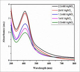
Figure 2. UV-Visible spectra of AgNPs obtained using water hyacinth leaves extract in different concentrations of AgNO3.
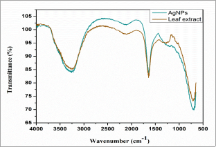
Figure 3. FTIR spectra of biosynthesized AgNPs and water hyacinth leaves extract.
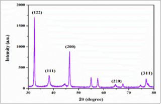
Figure 4. XRD pattern of biosynthesized AgNPs.
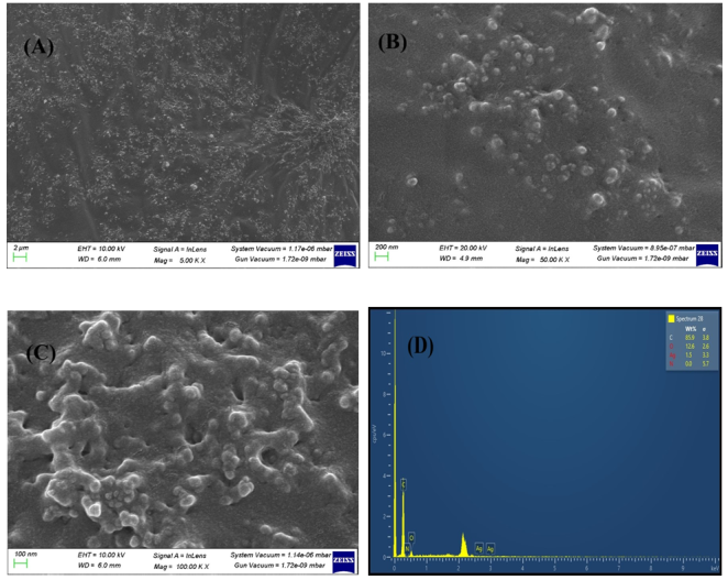
Figure 5. [A] [B] [C] SEM images showing synthesized AgNPs [D] EDAX image of synthesized AgNPs.
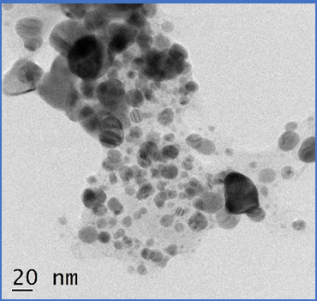
Figure 6. TEM image of synthesized AgNPs.
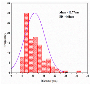
Figure 7. Particle size distribution histogram of AgNPs.
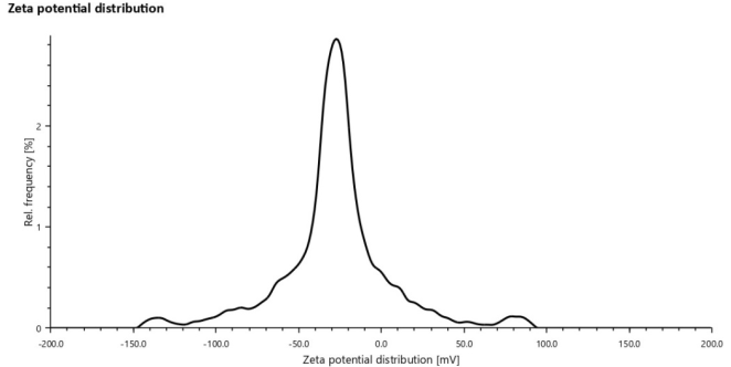
Figure 8. Zeta potential distribution of synthesized AgNPs.

Figure 9. Silver nanoparticles with the addition of metal ions after 10 minutes which was observed by UV-Visible spectra.
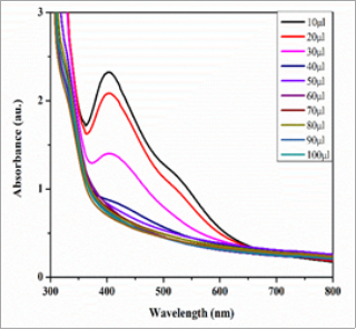
Figure 10. AgNPs UV-Visible spectra after the addition of different volumes of Fe3+ solution.
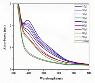
Figure 11. AgNPs UV-Visible spectra after the addition of different volumes of Hg2+ solution.